Showcase AM Expo 2018
Preparation for surgery in veterinary medicine
With the help of a 3D printed model from CT scans surgical intervention on a young horse with deformed hind legs was prepared. Thanks to optimal preparation the intervention was successful and the horse is now able to walk without problems.
-
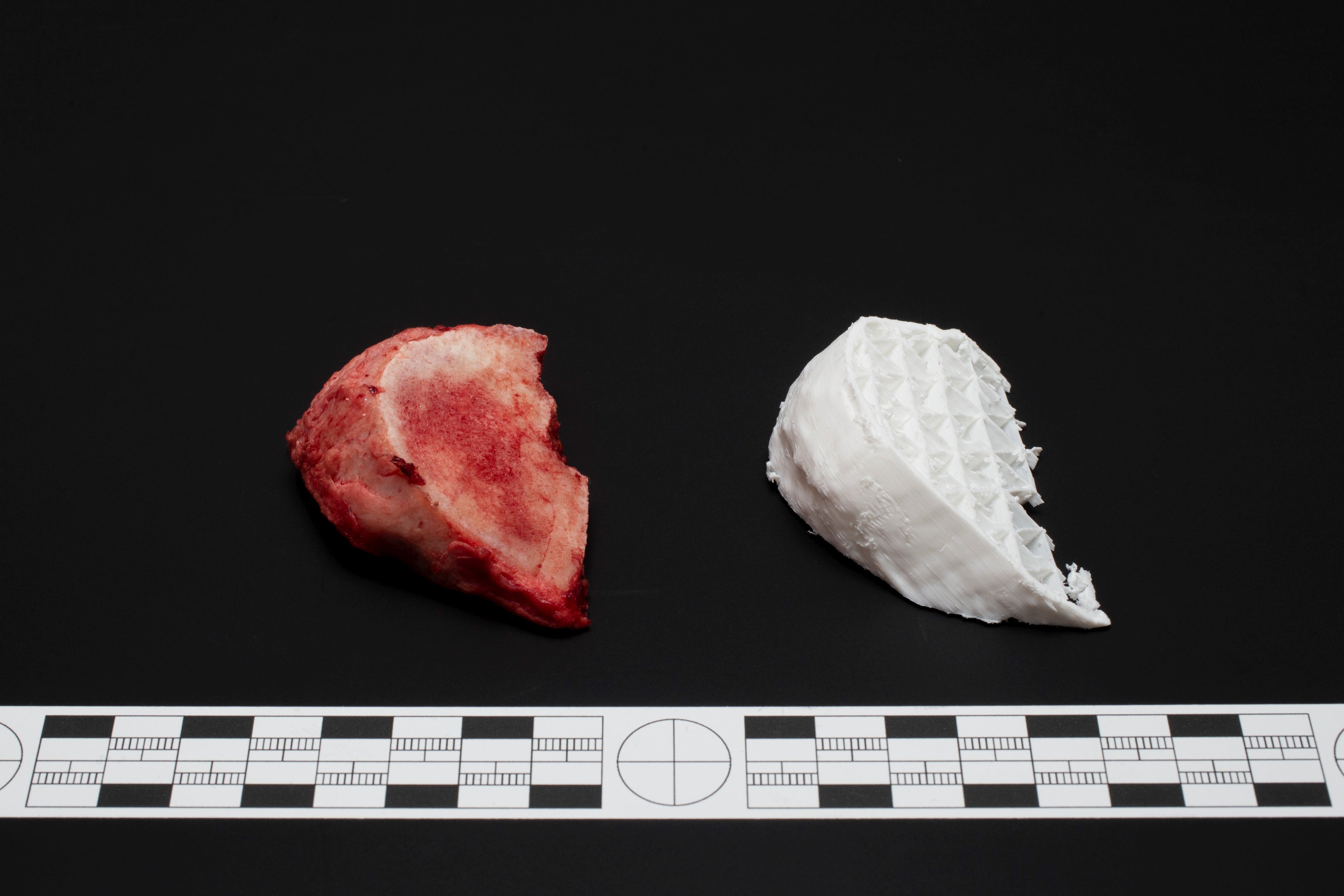
Cuts at the 3d printed model (right) and from surgery (left) -
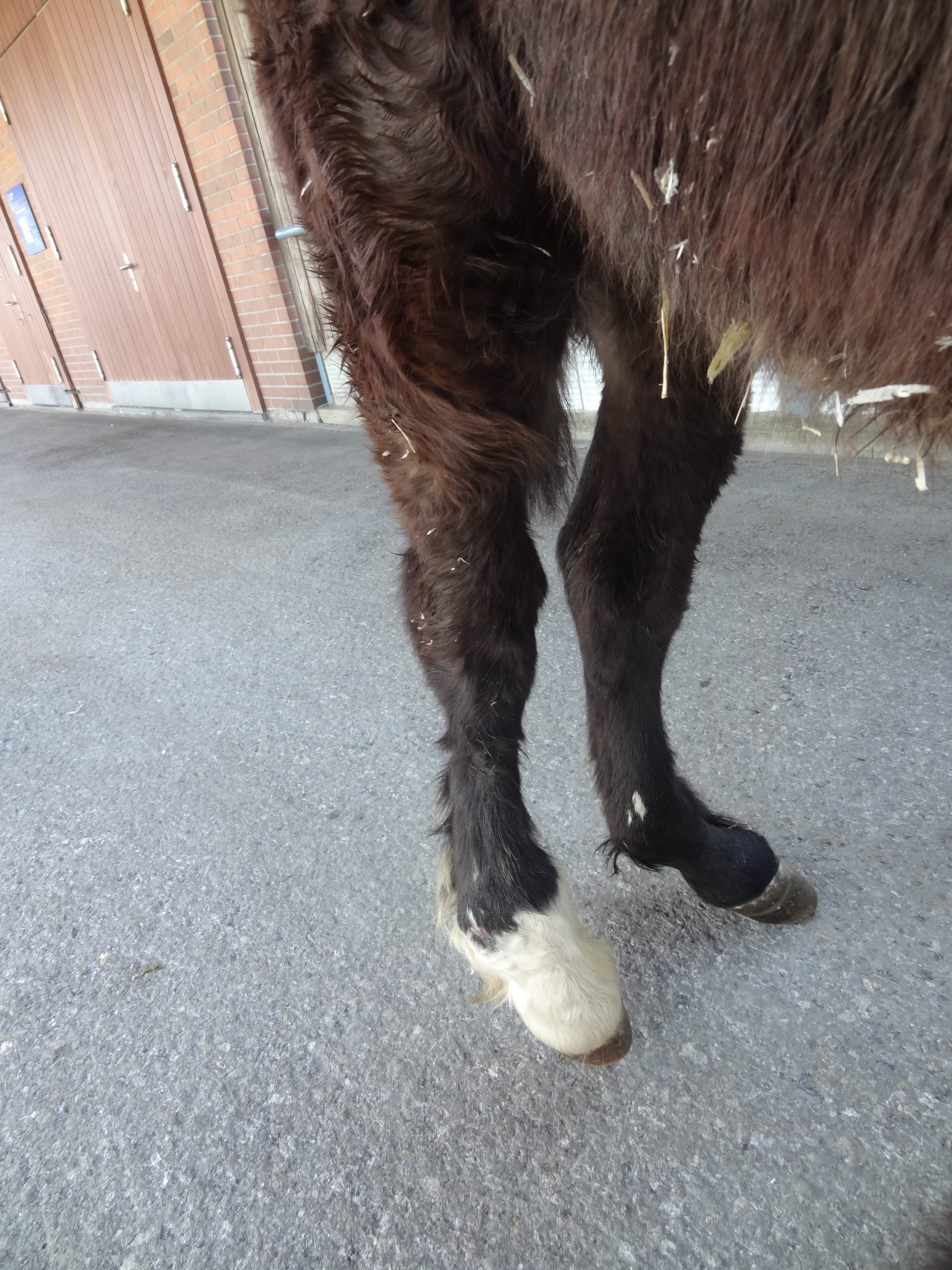
Horse before surgery with deformed hind legs -
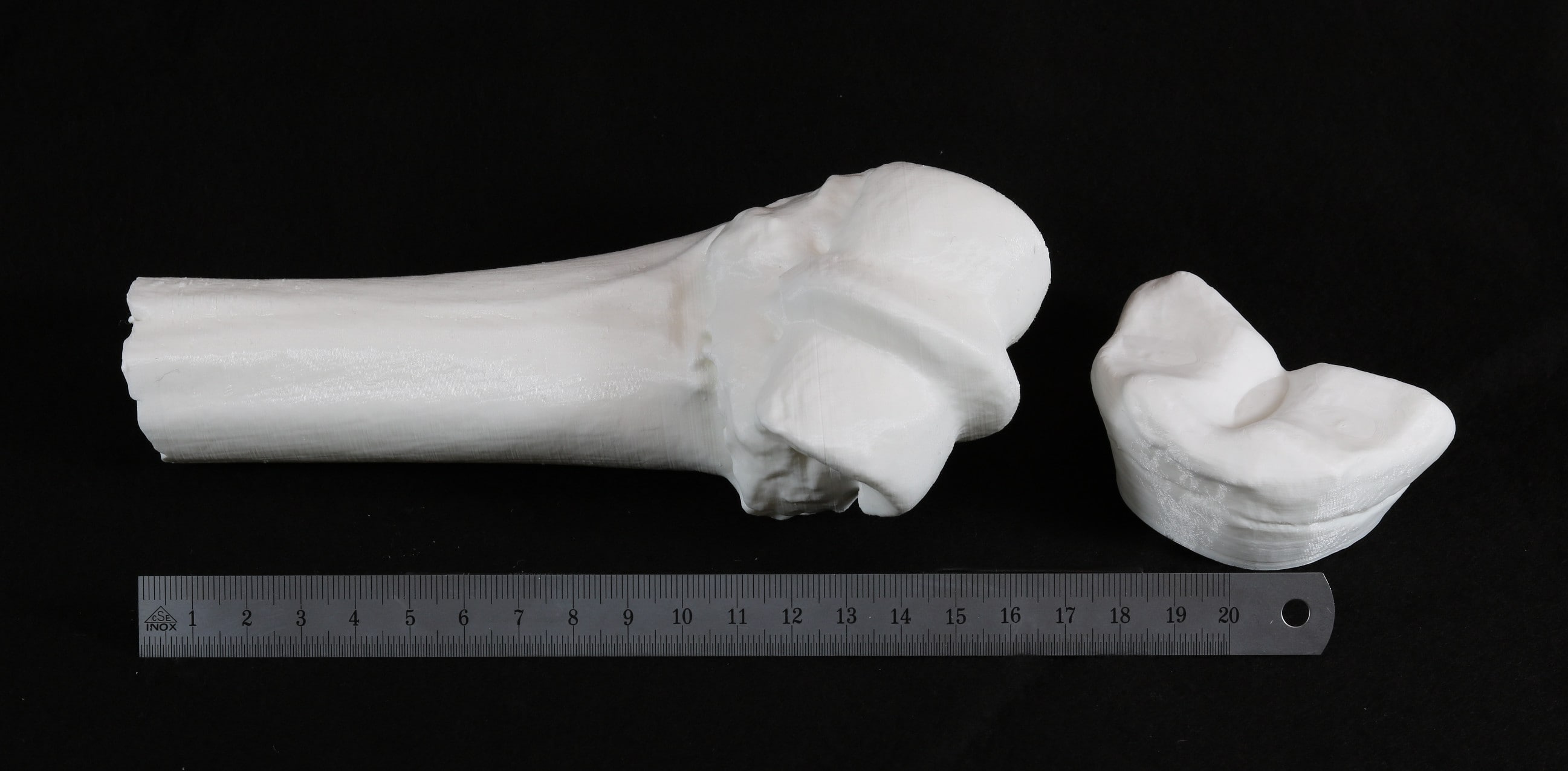
3D print of a leg bone -
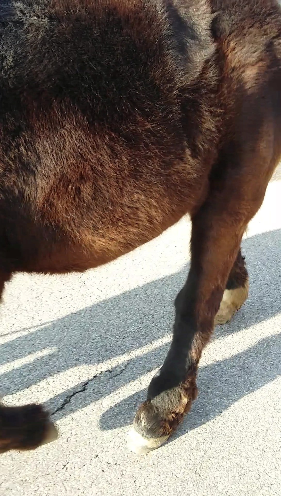
Horse with straight hind legs after surgery